
What Does Tooth Decay Look Like? Root and Hidden Decay Most People Never Picture
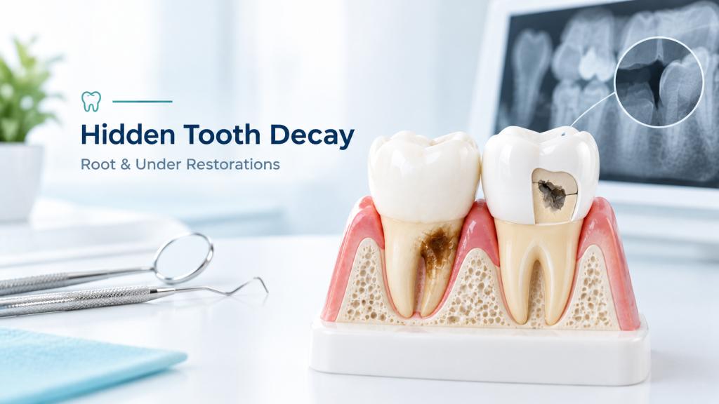
Most descriptions of tooth decay assume it’s happening on the visible, enamel-covered crown of a tooth, the part you see when you smile. Two of the most common forms of decay I treat don’t look anything like that picture. One happens below the gumline, on root surfaces never meant to face bacteria directly. The other happens underneath a filling or crown you already trusted to have solved the problem. Neither shows up as a classic white-to-brown spot on a visible tooth surface, which is exactly why both get missed so often.
Quick Answer
Root decay appears as a soft, leathery tan, brown, or black patch right at or just below the gumline, where gum recession has exposed the tooth root. Decay hiding beneath an old filling or crown often shows no visible surface change at all and is typically found only through an X-ray or a dentist’s probe catching a soft spot at the restoration’s edge. Both types are far more common in older adults and in anyone with existing dental restorations than the public tends to assume.
At a Glance
| Root Decay | Decay Under a Restoration | |
|---|---|---|
| Where it forms | At or below the gumline, on exposed root surface | Under the edge or floor of an existing filling or crown |
| What you’d see | A tan, brown, or black patch; texture feels leathery or soft | Often nothing visible; sometimes a dark line at the margin |
| How it’s usually found | Visual exam, gentle probing | X-ray, or a dentist’s explorer catching a soft spot |
| Who’s most affected | Older adults, anyone with gum recession | Anyone with older fillings or crowns, especially if margins have worn |
Root Decay: Why It Doesn’t Look Like a Regular Cavity
A cavity on the crown of a tooth starts in enamel, the hardest substance in the body, which is why it begins as a slow-forming white spot. Root decay starts somewhere completely different: on cementum and dentin, the softer tissues that make up the tooth root, tissues that were never designed to be exposed to the mouth’s bacteria and acid in the first place. Once gum recession or periodontal disease exposes that root surface, decay moves through it noticeably faster than it would through enamel, and it rarely bothers forming a neat white spot first. Instead, it tends to look tan, dark brown, or black almost from the start, with a soft, slightly leathery texture rather than a hard pit, right at the line where the tooth meets the gum.
This isn’t a rare or minor issue. According to data from the National Institute of Dental and Craniofacial Research, an estimated 42 percent of American adults aged 75 and older have untreated or restored root decay, and a separate national review of adults found untreated root decay specifically at around 10 percent, compared to 17.9 percent for untreated coronal (crown-surface) decay in the same population. As more people keep their natural teeth later in life, thanks to better prevention earlier on, more of them are living with exposed, vulnerable root surfaces for decades, which is exactly why this type of decay has become more common even as crown-surface cavities have declined.
Why Root Decay Targets Certain Patients So Specifically
Gum recession is the gateway, whether it comes from periodontal disease, years of aggressive brushing, or simply the gum tissue thinning with age. Dry mouth is just as significant a factor, since saliva neutralizes acid and clears away bacteria, and it decreases naturally with age while also being a common side effect of blood pressure medication, antidepressants, antihistamines, and dozens of other routinely prescribed drugs. Anyone managing a chronic illness that involves several daily medications is, often without realizing it, also managing a meaningfully higher root decay risk.
Hidden Decay: The Kind Forming Under Work You’ve Already Had Done
The second type is arguably sneakier, because there’s often genuinely nothing to see. Decay can develop underneath a filling or a crown, at the point where the restoration meets your natural tooth structure, a boundary dentists call the margin. Two closely related but distinct things can happen here, a distinction worth knowing because it isn’t just semantics: recurrent caries forms directly underneath the restoration itself, while secondary caries forms at the margin where the restoration meets the tooth. Both happen because bacteria find a way into microscopic gaps, whether from a restoration that’s worn down, a margin that was never perfectly sealed, or new decay forming independently at the edge of old work.
This is a genuinely major driver of restorative dentistry, not a footnote. Research published in the Journal of the Canadian Dental Association found that recurrent decay was the most frequent single cause of restoration failure in separate studies, accounting for 36 percent and 18.3 percent of failures respectively, and dental materials research more broadly points to replacing failed restorations as accounting for a substantial share of all restorative work performed. In plain terms, a meaningful portion of the fillings and crowns placed every year exist specifically to replace older restorations that failed this exact way.
What It Actually Looks (and Feels) Like
Visually, there’s often nothing at all, which is the whole problem. Occasionally a thin dark line appears right at the edge of a filling or along a crown’s margin, or the restoration’s color seems to have shifted slightly, but neither sign is reliable on its own. What actually reveals it is usually one of two things: an X-ray, which shows a dark shadow tucked beneath the denser, brighter restoration material the same way crown-surface decay shows up as a shadow, or a dentist’s exploratory instrument catching and sticking in a soft spot right at the margin during a routine exam, where a healthy margin would feel smooth and solid.
Why Neither of These Fits “What a Cavity Is Supposed to Look Like”
Most people’s mental image of decay, a visible white-then-brown-then-black spot that eventually becomes an obvious hole, applies reasonably well to decay on an exposed, enamel-covered tooth surface. Root decay skips the slow white-spot phase almost entirely because it’s working through softer tissue from the start. Decay under a restoration skips the visible phase altogether because it’s physically hidden by the filling or crown sitting on top of it. Both are legitimate tooth decay, both can progress to needing a root canal or extraction if ignored long enough, and neither one will show up if you’re only checking your teeth in the mirror.
Who Should Be Paying Closer Attention
If you’re over 60, have noticeable gum recession, take daily medications known to cause dry mouth, or have fillings or crowns that are more than 10 to 15 years old, you’re in the group where this guide matters most directly. None of that means something is currently wrong. It means routine X-rays and a dentist’s hands-on exam are doing real, specific work for you that a mirror simply can’t do.
What to Actually Do
Mention any of the risk factors above to your dentist directly, rather than assuming they’ll automatically investigate without being prompted, particularly dry mouth from medication, which patients often don’t think to bring up in a dental context. Keep up with the X-ray schedule your dentist recommends, since this is specifically how hidden and root decay get caught before they progress. And if a tooth with an old filling or crown develops new sensitivity to temperature or pressure, treat that as worth a dental visit even though there’s nothing to see, since sensitivity is frequently the only symptom either of these produces before an X-ray confirms what’s happening underneath.
When It’s Gone Too Far to Restore
Root decay and recurrent decay under old restorations are two of the more common reasons a tooth that seemed stable for years suddenly isn’t. If decay has progressed to the point where a tooth can no longer support a filling or a new crown, that opens the extraction-versus-save-it conversation. Our guide to root canal plus crown versus implant cost walks through how that decision gets made, and our dental implants cost guide covers what replacement involves if saving the tooth isn’t realistic.
Frequently Asked Questions
Is root decay more serious than a regular cavity?
It can progress faster because it’s working through softer root tissue rather than enamel, and it’s more likely to go unnoticed since it rarely forms the early white-spot stage that crown-surface decay does. Both types need professional treatment either way.
Can decay under a crown happen even if the crown looks and feels perfectly fine?
Yes. This is exactly what makes it hard to catch. A crown can look and feel completely normal on the surface while decay develops at the margin underneath it, which is why routine X-rays matter even for teeth that already have restorative work.
What’s the real difference between recurrent and secondary caries?
Recurrent caries forms directly beneath the restoration. Secondary caries forms at the margin where the restoration meets the natural tooth. Both stem from bacteria finding a way past the restoration’s seal, and both are treated similarly, but the distinction matters for how a dentist plans the repair.
Does gum recession always lead to root decay?
No, but it’s the necessary first step, since root decay can’t form on a surface that’s still covered by healthy gum tissue. Recession simply creates the exposure that makes root decay possible if other risk factors, like dry mouth or reduced hygiene access in that area, are also present.
Can root decay be prevented once recession has already happened?
Fluoride, addressing dry mouth directly with your physician or dentist, and careful hygiene specifically at the gumline all meaningfully reduce the risk, even though the exposed root surface itself can’t be reversed.
How often should X-rays be taken if I have several old fillings or crowns?
This depends on your personal risk profile, which your dentist determines, but a higher number of existing restorations is itself a reason some patients are recommended more frequent imaging rather than less.
The Bottom Line
Tooth decay doesn’t only show up as the slow white-to-black progression most people picture on a visible tooth surface. Root decay moves faster through softer tissue right at the gumline and skips the early warning stage almost entirely. Decay under an old filling or crown often shows nothing on the surface at all. Both are common, both are specifically why dentists rely on X-rays and hands-on exams rather than a visual check alone, and both are worth asking about directly if you fall into one of the higher-risk groups above.
Sources: National Institute of Dental and Craniofacial Research, Tooth Loss Data and Statistics · Carpendale, Restoration of the Failing Dentition, Journal of the Canadian Dental Association